Why tissue section quality matters
In life sciences and biomedical research, every breakthrough depends on one critical factor: the quality of tissue sections prepared for analysis. Whether the focus is the brain, heart, lungs, liver, kidneys or plant tissues, poor sectioning can compromise even the most advanced imaging and analytical systems.
Vibrating microtomes have become indispensable because they are specifically engineered to preserve tissue integrity while producing precise, reproducible sections that closely resemble the tissue’s natural state.
What is a vibrating microtome?
A vibrating microtome is a precision instrument designed to section fresh, unfixed or lightly fixed biological tissues without paraffin embedding or freezing. Instead of cutting through hardened blocks, the instrument uses a vibrating blade combined with controlled forward motion to gently slice delicate specimens.
This approach significantly reduces mechanical stress and helps preserve:
- Cellular architecture
- Neural networks
- Tissue morphology
- Physiological viability
The result is a high‑quality tissue section that is ideal for advanced biological and medical research.
Why tissue preservation is critical
Every tissue sample carries valuable biological information. If sectioning damages cells, tears fine structures or introduces artifacts, essential data may be lost before the experiment even begins.
High‑quality sectioning enables researchers to:
- Preserve natural tissue architecture
- Maintain cell viability
- Reduce mechanical damage
- Improve imaging quality
- Increase experimental reproducibility
- Generate more reliable scientific results
In many applications, the quality of the slice directly determines the quality of the science.
How a vibrating microtome works
Step 1: Tissue preparation
Fresh tissue is carefully mounted onto the specimen holder using appropriate support media to stabilise and orient the sample for sectioning.
Step 2: Precision vibration
A finely engineered blade oscillates at a controlled frequency while advancing through the specimen. The vibration minimises cutting resistance and reduces deformation instead of forcing the blade through tissue.
Step 3: Controlled sectioning
Researchers can precisely adjust parameters such as:
- Blade oscillation frequency
- Cutting amplitude
- Advance speed
- Section thickness
These controls allow optimisation for different tissue types and experimental protocols.
Step 4: Collection
Freshly cut sections are collected immediately for downstream applications, including microscopy, electrophysiology, molecular biology and live‑cell imaging.
Where vibrating microtomes are used
Vibrating microtomes support a wide range of research disciplines and model systems across neuroscience, organ physiology, plant biology and advanced translational research.
Key application areas
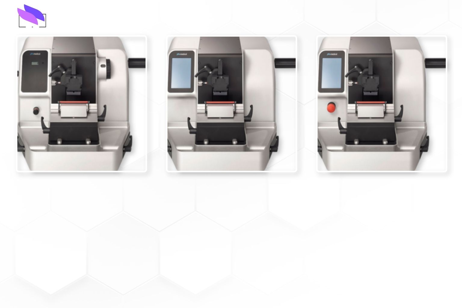
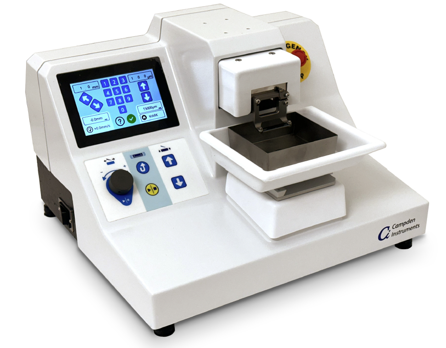
What makes modern vibrating microtomes different?
Contemporary instruments combine mechanical precision with intuitive control systems, helping laboratories achieve consistent results across users and studies.
Key design and workflow features include:
- Intuitive touchscreen interfaces and clear parameter displays
- Programmable slicing protocols for repeatable methods
- Real‑time monitoring of cutting speed and vibration
- Integrated or optional cooling to protect sensitive tissues
- User‑updatable firmware for ongoing performance improvements
- Modular, serviceable designs that reduce downtime
Precision that supports better science
A defining strength of modern vibrating microtomes is their ability to maintain blade stability and cutting accuracy while working with soft, delicate material. Combined with fine control of advance speed and vibration settings, this gives researchers the flexibility to work confidently across a wide variety of tissue types.
This level of precision supports:
- Better preservation of tissue integrity
- Improved experimental consistency between batches
- Reduced sample loss and repeat sections
- Greater confidence in downstream measurements and conclusions
Supporting the future of biomedical research
As research advances in personalised medicine, regenerative therapies, neuroscience and molecular diagnostics, demand for high‑quality tissue preparation will continue to rise.
Vibrating microtomes have evolved from niche tools into enabling technologies that help laboratories generate accurate, reproducible and clinically relevant data. Each well‑prepared section contributes to deeper biological insight and ultimately supports better diagnostic and therapeutic solutions.
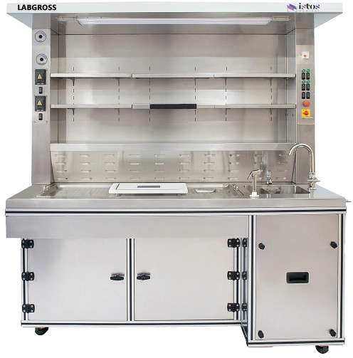
ISTOS Medical: enabling precision tissue sectioning
At ISTOS Medical, we support researchers, universities, medical institutions, pharmaceutical companies and life science laboratories with advanced histopathology and research instrumentation designed for precision, reliability and innovation.
To explore vibrating microtome solutions or broader histopathology workflows, visit www.istosmedical.com .
Because every discovery begins with a perfectly prepared sample.