Introduction
When people think about histopathology, they often picture microscopes, tissue samples and pathology reports. Yet behind every accurately diagnosed specimen lies a sophisticated blend of mechanical engineering, electronics, automation and precision manufacturing working quietly in the background.
Modern tissue processors, microtomes, cryostats, embedding centers, slide stainers, coverslippers and grossing stations are no longer just simple machines. They are intelligent systems powered by advanced motherboards, microprocessors, sensors and software-driven control, designed to deliver consistent and reproducible results case after case.
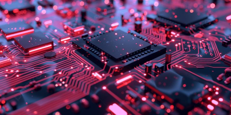
The Motherboard: The Brain of Histopathology Instruments
Just as the human brain coordinates complex biological functions, the motherboard serves as the central control unit of many histopathology instruments. It orchestrates how mechanical, thermal, electrical and software subsystems work together to process tissue safely and efficiently.
A modern instrument motherboard typically integrates:
- Microprocessors (CPU) for real-time control and decision-making
- Memory modules for storing programs, logs and configuration data
- Power management circuits to ensure stable operation of all components
- Communication interfaces for peripherals, networks and service tools
- Sensor controllers to read temperature, pressure and position feedback
- Motor drivers to control stepper, servo and DC motors
In an automated tissue processor, for example, the motherboard manages reagent changes, processing schedules, vacuum cycles, temperature regulation and safety monitoring with minimal operator intervention, ensuring consistent processing every day.
Embedded Microcontrollers and Intelligent Control
At the heart of many histopathology devices are embedded microcontrollers and dedicated control boards. These compact computing units execute specific tasks with high reliability and are optimized for medical device environments.
Typical responsibilities of embedded control systems include:
- Continuous temperature monitoring and regulation
- Motion control for specimen movement and cutting mechanisms
- Sensor feedback interpretation for safety and performance
- Alarm detection and management for fault conditions
- User interface operation via keypads or touchscreens
- Data logging for traceability and quality management
Whether stabilizing a cryostat chamber at sub-zero temperatures or maintaining precise paraffin temperatures in an embedding center, embedded electronics ensure consistency, repeatability and adherence to laboratory protocols.
Precision Motors: The Unsung Heroes of Motion
Mechanical movement is essential to histopathology workflows, from advancing a tissue block in a microtome to moving slides through automated stainers and coverslippers. This motion is made possible by carefully engineered drive systems.
Many laboratory instruments utilize:
- Stepper motors for controlled, incremental motion
- Servo motors for precise, feedback-driven positioning
- Linear actuators for straight-line motion of platforms and carriages
- Gear assemblies for torque transmission and speed reduction
- Precision bearings for smooth, low-friction movement
In a microtome, for example, the specimen may advance only a few microns with each cutting cycle. Achieving this level of precision requires finely tuned mechanical components working in harmony with electronic control systems. Even small deviations can affect section thickness, morphology and ultimately diagnostic interpretation.
Sensors Safeguarding Sample Integrity
Histopathology workflows rely heavily on stable environmental conditions and safe operation of equipment. Modern instruments incorporate a range of sensors to continuously monitor critical parameters and protect both samples and users.
Commonly used sensors include:
- Temperature sensors for reagent, paraffin and chamber control
- Humidity sensors in climate-sensitive environments
- Position sensors for doors, lids and moving stages
- Pressure sensors for vacuum and pneumatic systems
- Liquid level sensors for reagents and wax reservoirs
- Safety interlock sensors to prevent unsafe operation
For example, a tissue processor must maintain precise temperatures and reagent levels throughout the entire processing cycle to preserve tissue morphology and prevent incomplete infiltration. Sensor feedback ensures that any deviation is detected and corrected promptly.
Software and Hardware Working in Harmony
Today’s histopathology instruments are increasingly software-driven. User interfaces, configuration tools and diagnostic logs are all managed through embedded software that sits on top of the mechanical and electronic layers.
Advanced systems often provide:
- Touchscreen interfaces for intuitive operation
- Workflow automation and guided protocols
- On-screen error diagnostics and troubleshooting
- User access management and audit trails
- Data recording for quality and compliance
- Remote monitoring and service access
By integrating software with mechanical and electronic systems, laboratories can improve productivity, reduce manual errors and maintain consistent quality standards across high workloads and multiple operators.
Cryostats: A Showcase of Engineering Integration
Cryostats represent one of the most sophisticated combinations of mechanical and electronic engineering found in histopathology. They are essential for frozen section diagnostics, especially during intraoperative consultations.
A typical cryostat integrates:
- Refrigeration technology to achieve and maintain sub-zero temperatures
- Precision temperature control systems for stability and uniformity
- Electronic sensors to monitor chamber and specimen conditions
- Microprocessor-based controllers for cycle logic and safety
- High-precision microtomes for frozen tissue sectioning
The result is the ability to rapidly prepare frozen tissue sections with high-quality morphology, enabling timely diagnostic decisions during surgery while maintaining patient safety and workflow efficiency.
The Future: Smarter Histopathology Laboratories
The next generation of histopathology equipment is becoming increasingly intelligent and connected. Laboratories are moving toward digital-ready, data-rich environments where instruments communicate with information systems and decision-support tools.
Emerging innovations include:
- AI-assisted diagnostics and pattern recognition
- Predictive maintenance based on equipment usage and performance data
- IoT connectivity for real-time status and alerts
- Digital pathology integration and image-based workflows
- Cloud-based monitoring and reporting
- Automated workflow optimization across instrument platforms
As laboratories continue to modernize, the importance of reliable electronics, advanced motherboards, precision mechanics and intelligent automation will only continue to grow.
More Than Just Machines
Every motherboard, sensor, chip, motor and mechanical component inside a histopathology instrument ultimately serves a single purpose: supporting accurate diagnosis and better patient outcomes. While pathologists interpret slides and clinicians make treatment decisions, the unseen engineering inside laboratory equipment ensures that tissue samples are processed, sectioned, stained and prepared with the precision required for confident diagnosis.