Strengthening Histopathology Across Maharashtra
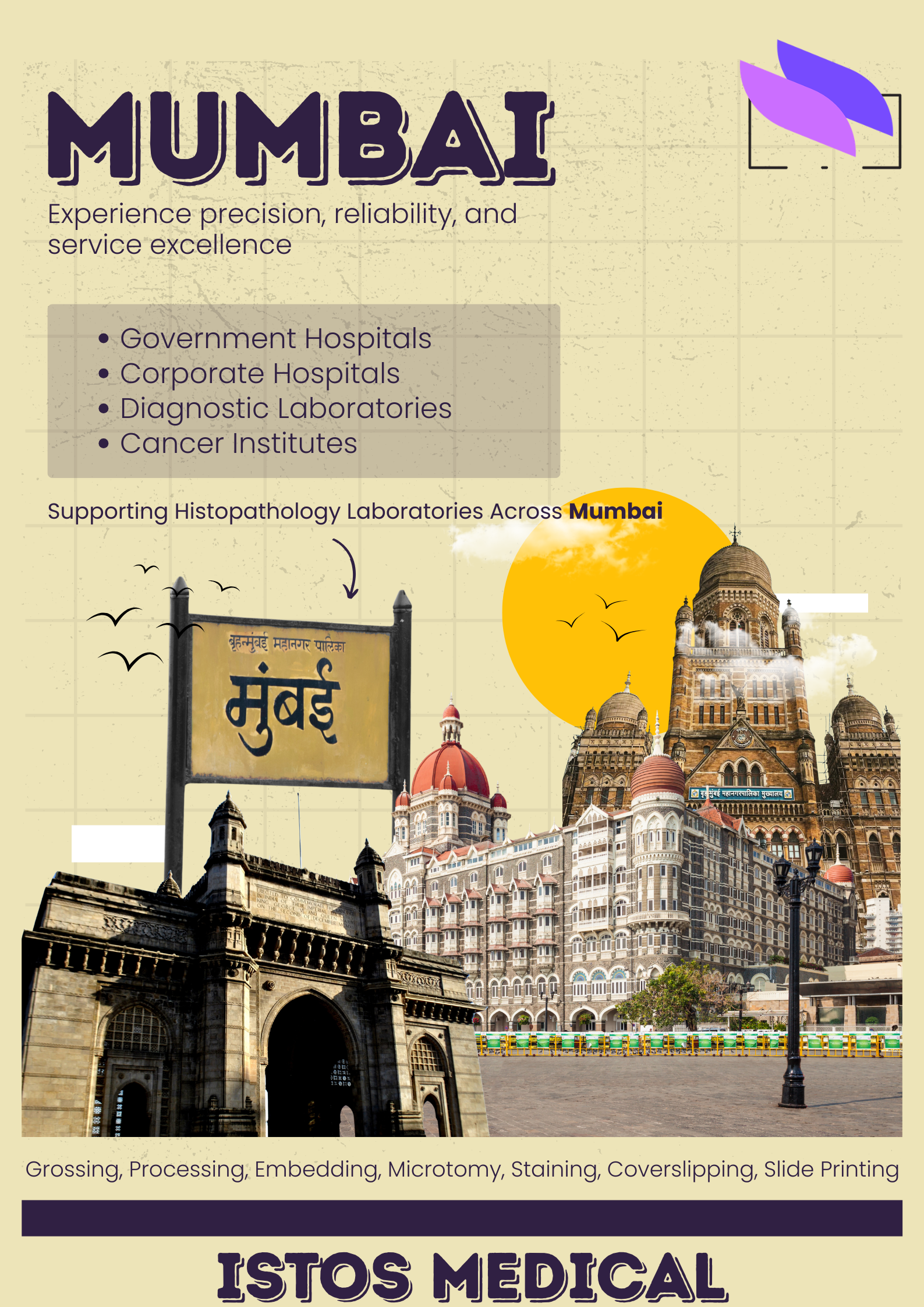
Supporting Better Diagnostics
Maharashtra is home to some of India’s most respected government hospitals, corporate healthcare networks, cancer institutes and diagnostic laboratories. Every day, thousands of biopsies are processed across this ecosystem, and each specimen depends on a reliable histopathology workflow.
This refactored template is designed to showcase how ISTOS Medical supports laboratories across Maharashtra with a cleaner layout, wider reading width and improved tables—while keeping the same familiar look and feel as your existing blog design.
In the sections that follow, we highlight hospital partners, explore the state’s healthcare strengths, outline histopathology workflow solutions and share practical recommendations for different types of laboratories across Maharashtra.
Hospital Highlights
ISTOS Medical collaborates with a wide range of hospitals and institutes, from teaching medical colleges to tertiary cancer centres. The following spotlight cards illustrate the diversity of clinical environments where dependable histopathology technology plays a central role.
BJ Medical College, Pune
A high‑volume teaching institution where histopathology supports undergraduate and postgraduate training, daily diagnostic work and multi‑disciplinary tumour boards.
Reliable section quality and well‑designed grossing areas help faculty maintain consistent standards across large caseloads and diverse case types.
LTMG Sion Hospital, Mumbai
A major government hospital serving a dense urban catchment, with histopathology at the core of surgical and oncology services.
High daily biopsy volumes make equipment robustness, ease of maintenance and predictable turnaround time particularly important.
Tata Memorial Hospital, Mumbai
A leading cancer centre where complex resections, core biopsies and specialised stains demand exceptional section quality and workflow reliability.
Histopathology technology supports not only routine diagnostics, but also clinical research, tumour boards and advanced oncology programmes.
More as to Why Maharashtra Leads Healthcare in India
Metropolitan clusters such as Mumbai, Navi Mumbai and Pune anchor advanced oncology, transplant and multi‑specialty services, while growing cities like Nagpur, Nashik and Aurangabad steadily expand diagnostic capacity.
This breadth makes Maharashtra a reference point for histopathology services in western India. For ISTOS Medical, it also means working with laboratories at multiple stages of development—from compact district hospitals upgrading their first microtome to large corporate chains standardising workflows across campuses.
Key Drivers of Histopathology Demand
- Rising oncology caseloads requiring timely biopsy interpretation and margin assessment.
- Growth in minimally invasive procedures that depend on accurate tissue diagnosis.
- Expansion of day‑care surgeries and short‑stay units, increasing routine section volumes.
- Greater emphasis on quality assurance, accreditation and laboratory safety standards.
- Emerging adoption of digital pathology and telepathology for remote case review.
As these drivers converge, laboratories across Maharashtra look for partners who can provide integrated histopathology solutions rather than isolated instruments. A joined‑up approach helps them manage rising workloads without compromising diagnostic confidence.
Maharashtra Pathology Snapshot
This landscape creates both opportunity and responsibility: laboratories must deliver timely, accurate results while operating within real‑world constraints such as limited bench space and staffing.
Government Hospitals & Teaching Institutions
Government hospitals and medical colleges in Maharashtra carry a dual mandate: provide accessible care to large populations and train the next generation of clinicians and pathologists. Their histopathology departments handle everything from routine biopsies to complex resections, often within tight budget and infrastructure boundaries.
In these environments, ISTOS Medical focuses on durable, service‑friendly technology that can withstand continuous daily use. Equipment is selected not only for performance, but also for ease of training, safety and straightforward maintenance.
- BJ Medical College and similar teaching centres where trainees learn tissue processing and sectioning techniques.
- Large municipal hospitals that serve as referral hubs for surrounding districts.
- District hospitals upgrading from fully manual to semi‑automated histopathology workflows.
By aligning equipment choices with teaching needs, ISTOS helps ensure that new pathologists and technologists become comfortable with modern histopathology tools early in their careers. This supports smoother adoption of advanced systems in the future.
Corporate & Multispecialty Hospitals
Corporate hospitals and multispecialty networks place a strong emphasis on standardisation, patient experience and integrated workflows. Their histopathology laboratories often serve multiple campuses, day‑care centres and speciality units, making consistency and scalability essential.
In these settings, ISTOS Medical emphasises harmonised equipment portfolios: microtomes, processors, embedding centres and stainers that share similar user interfaces and operating principles. This simplifies training and helps technologists move between units with minimal disruption.
- Standardised section quality across multiple hospital sites.
- Clear, traceable specimen journeys from theatre to histology report.
- Equipment layouts designed to fit high‑density urban campuses.
- Service and support structures that minimise downtime and protect theatre schedules.
For growing networks, ISTOS can also help plan phased upgrades, ensuring that new systems integrate smoothly with existing laboratory information systems and reporting tools.
Cancer Institutes & Oncology Labs
In oncology, histopathology is central to staging, grading and treatment planning. Dedicated cancer hospitals and oncopathology laboratories in Maharashtra rely on precise sectioning, reliable frozen section support and well‑controlled staining workflows.
ISTOS Medical’s histopathology solutions in these environments focus on repeatable precision: stable rotary microtomes for delicate tissue, cryostats for intra‑operative decisions, and staining systems that support both routine H&E and more specialised immunohistochemistry protocols.
As tumour boards and multi‑disciplinary clinics become more common, oncology laboratories also look towards digital workflows. Well‑prepared sections and high‑quality slides are the foundation for accurate scanning, image sharing and collaborative review.
Diagnostic Laboratories & Regional Pathology Centres
Independent diagnostic laboratories and regional pathology centres complement hospital‑based services by handling routine biopsies, referral cases and community screening programmes. Many operate as hubs for satellite collection centres across Maharashtra.
For these laboratories, ISTOS Medical recommends modular equipment configurations that can grow with demand. Compact grossing areas, reliable tissue processors and versatile microtomes form a solid starting point, with room for future automation and digital integration.
- Smaller laboratories beginning with core histopathology capabilities.
- Regional centres steadily increasing surgical pathology volumes.
- Networks planning eventual digital pathology adoption for remote reporting.
When volumes rise, laboratories can progressively add automated stainers, slide printers and scanner‑ready workflows, ensuring that each upgrade builds on existing strengths rather than forcing disruptive overhauls.
Histopathology Workflow
A strong histopathology service depends on the quality of each step in the workflow. ISTOS Medical supports laboratories by looking at the process end‑to‑end—from specimen reception and grossing through processing, embedding, sectioning, staining and slide identification.
| Stage | Requirement | Solution |
|---|---|---|
| Grossing | Safe, organised specimen handling with adequate space, lighting and ventilation. Technologists need ergonomic work areas that support both simple biopsies and complex resections. | Grossing stations designed specifically for histopathology workflows, incorporating ventilation, lighting, sink arrangements and waste management. |
| Processing | Consistent dehydration, clearing and paraffin infiltration to preserve tissue morphology and minimise artefacts. | Tissue processors offering reliable, repeatable programs, suitable for routine and high‑throughput environments. |
| Embedding | Correct orientation of tissue in blocks, stable temperature control and comfortable working posture for technologists. | Embedding centres with thermostatically controlled paraffin, cooling plates and intuitive layouts to reduce fatigue. |
| Sectioning | Thin, uniform sections with minimal compression and chatter, adjustable for different tissue types and diagnostic needs. | Rotary microtomes and cryostats with smooth mechanics and fine‑tuned controls for thickness, cutting speed and specimen advance. |
| Staining | Reliable stain consistency, cleaner slide presentation and reduced manual handling to protect tissue and improve safety. | Slide stainers and coverslippers configured for routine H&E and special stains, with options for more advanced protocols. |
| Identification | Clear, durable slide and cassette labelling to support traceable case flow and minimise identification errors. | Slide printers and labelling systems integrated into the overall workflow, improving traceability from grossing to reporting. |
Regional Presence Across Maharashtra
ISTOS Medical’s work in Maharashtra spans major metropolitan centres and fast‑growing regional hubs. From Mumbai and Navi Mumbai to Pune, Nashik, Nagpur, Aurangabad and suburban belts such as Mira Road, Borivali, Ghatkopar, Hadapsar and Pimpri, laboratories use histopathology technology to support diverse clinical programmes.
Urban campuses often require compact, high‑capacity configurations that fit limited space, while district centres prioritise multi‑purpose workstations and straightforward maintenance. By understanding these local needs, ISTOS designs solutions that feel at home in each environment.
The goal is simple: help ensure that high‑quality histopathology services are available where patients live and seek care, not only in a few flagship institutions.
Recommended Configurations by Laboratory Type
Although every laboratory is unique, certain equipment combinations tend to work particularly well in specific segments. The table below outlines typical starting configurations and upgrade paths for different types of histopathology services across Maharashtra.
| Laboratory Type | Recommended Configuration | Upgrade Path |
|---|---|---|
| Teaching & Government Hospitals | Manual or semi‑automated rotary microtomes, robust grossing stations and reliable tissue processors suitable for high daily workloads and training. | Gradual introduction of automated stainers, enhanced ventilation and digital‑friendly slide preparation as caseloads and resources grow. |
| Corporate & Multispecialty Hospitals | Standardised microtome models across sites, mid‑ to high‑capacity tissue processors, embedding centres and stainers with similar user interfaces. | Expansion to include slide printers, integrated LIS connectivity and scanner‑ready staining workflows, supporting future digital pathology adoption. |
| Cancer Institutes & Oncopathology Centres | High‑precision microtomes, cryostats for intra‑operative margin assessment and stain systems tuned for specialised oncology protocols. | Deployment of digital workstations for tumour boards, remote case review and multi‑disciplinary planning conferences. |
| Independent Diagnostic Laboratories | Compact grossing, processing and sectioning setups focusing on reliability and ease of use, ideal for routine biopsy volumes. | Stepwise addition of automation, slide labelling and digital slide preparation as referral numbers and service offerings expand. |
| Research & Academic Centres | Versatile combinations capable of handling routine histology, experimental protocols and teaching needs, including frozen sections when required. | Introduction of digital slide archives, image analysis tools and collaborative platforms to support long‑term studies and training programmes. |