Introduction
In Histopathology, formaldehyde is most often used as 10% Neutral Buffered Formalin (NBF). It is the standard fixative that “locks” tissue soon after surgery or biopsy so that the microscopic appearance reflects the patient’s true pathology rather than post‑removal changes.
1. Preserving Tissue Structure
Once tissue leaves the body, cells start to break down and lose their natural architecture. Enzymes, bacteria and physical handling can quickly destroy critical details unless the specimen is stabilised.
- Stops self‑digestion of cells by inactivating enzymes.
- Maintains overall cell and tissue architecture.
- Prevents decomposition during storage and transport.
- Helps ensure that what the pathologist sees resembles the situation in vivo.
Good fixation is the difference between a crisp, interpretable slide and one that leaves too much room for doubt.
2. Protein Cross‑linking and Structural Stability
Formaldehyde works by forming chemical bridges between proteins inside the tissue. These cross‑links hold cell structures in place so they can withstand processing, cutting and staining without collapsing or washing away.
- Stabilises nuclei and cytoplasm so that cell outlines remain clear.
- Reduces distortion and tearing when the tissue is dehydrated and embedded.
- Supports smooth, consistent sectioning at the microtome.
- Creates a stable platform for special stains and immunohistochemistry.
When fixation is poor, tissues can be soft, friable and difficult to cut, and fine details that matter for diagnosis may be lost.
3. Controlling Microbial Growth
Fresh surgical and biopsy material is an ideal nutrient source for bacteria and fungi. If microbes are allowed to grow, they can damage tissue and compromise staining.
- Reduces bacterial and fungal overgrowth in stored specimens.
- Helps maintain sample integrity during longer transport times.
- Supports cleaner, more reproducible slides in routine workloads.
This antimicrobial effect is particularly important when specimens travel from distant collection sites to central laboratories.
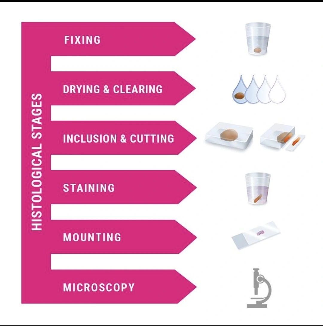
4. Foundation for the Tissue Processing Workflow
Fixation is the first major step before tissue enters a processor, embedding centre, microtome or stainer. If that first step is weak, every downstream stage has to fight against it.
Typical problems linked to inadequate fixation include:
- Incomplete infiltration with paraffin and processing artefacts.
- Poor or uneven staining, especially in deeper tissue levels.
- Tissue shrinkage, cracking or excessive hardness.
- Microtome chatter, folds, knife marks and torn sections.
- Slides that are technically acceptable but diagnostically frustrating.
In practice, many “staining” or “instrument” issues are in fact fixation problems that show up late in the workflow.
5. Impact on Staining Quality
Routine stains such as Hematoxylin & Eosin (H&E), PAS, Masson’s Trichrome and a wide range of IHC markers all assume that the tissue has been fixed in a controlled, reproducible way.
- Sharper nuclear and cytoplasmic detail on H&E.
- More predictable uptake of special stains.
- Better antigen preservation and signal‑to‑noise for IHC.
- Greater confidence that differences between slides are biological, not technical.
When fixation varies from case to case, staining becomes harder to standardise and subtle diagnostic features are easier to miss.
Recommended Fixation Times in 10% NBF
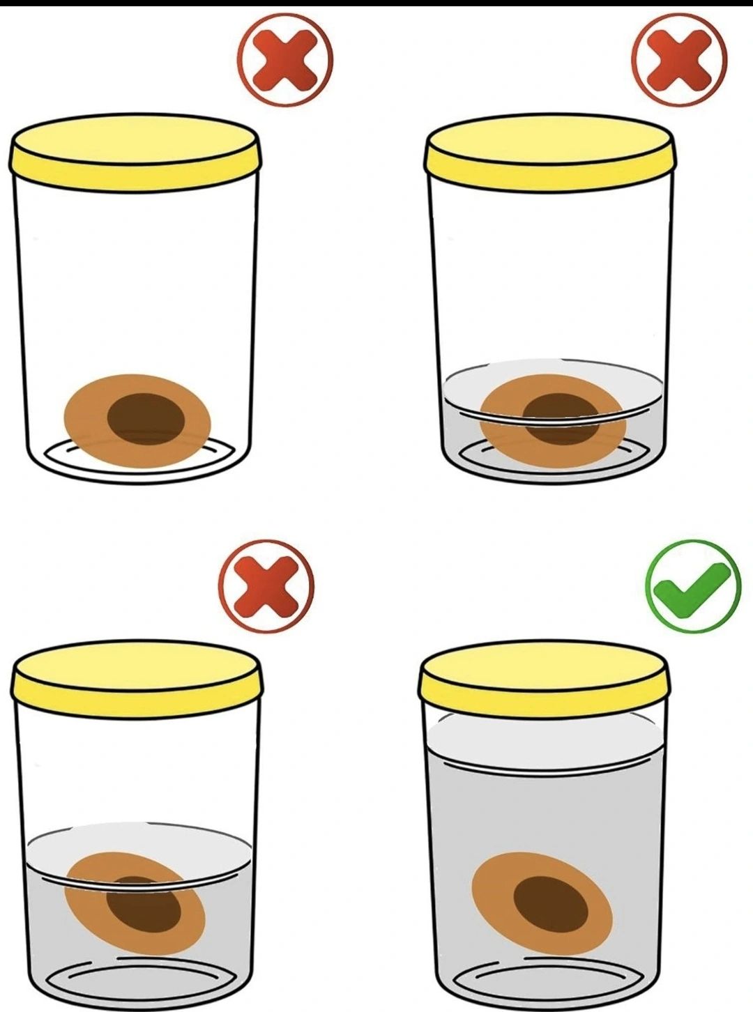
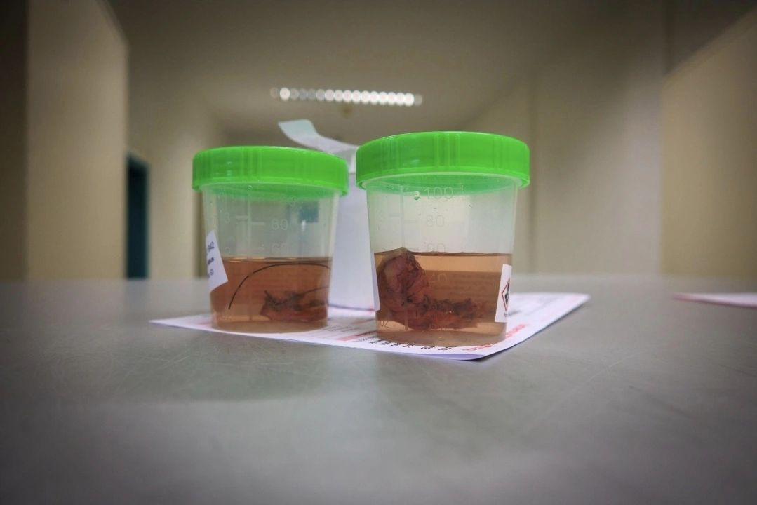
Ten percent Neutral Buffered Formalin contains roughly 4% formaldehyde in a buffered aqueous solution. Exact fixation times depend on the size and type of specimen, but most routine workflows follow simple size‑based ranges.
- Small biopsies: typically around 6–12 hours.
- Routine surgical specimens: typically around 12–24 hours.
- Large resections: often 24–48 hours, with appropriate trimming for penetration.
Under‑fixation risks poor morphology and inconsistent staining; over‑fixation can make tissue brittle and may mask some epitopes. The goal is controlled, documented times rather than “as long as it takes”.
Safety and Formaldehyde Exposure
Formaldehyde is an irritant and potential sensitiser, so laboratories must treat it as a controlled chemical rather than a routine commodity. Good engineering controls and work practices protect both staff and the surrounding environment.
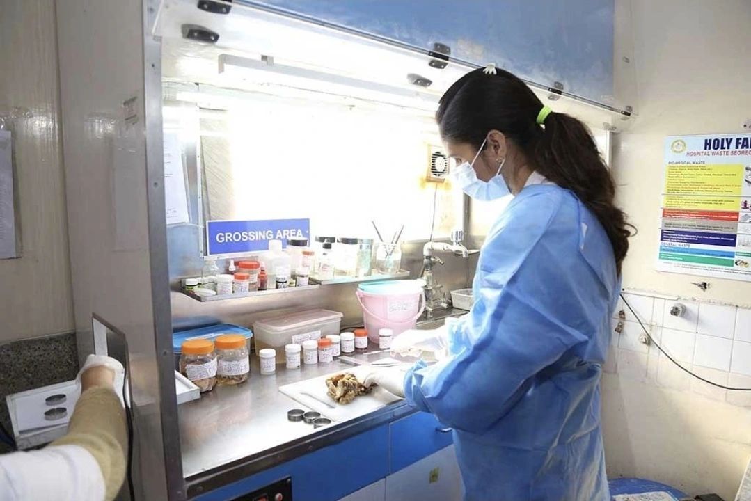
- Use grossing benches with local extraction to capture vapours at the source.
- Wear appropriate PPE: gloves, eye protection, lab coats and respiratory protection where indicated.
- Handle and store formalin in clearly marked containers and designated chemical storage areas.
- Train staff in spill management, neutralisation and safe disposal procedures.
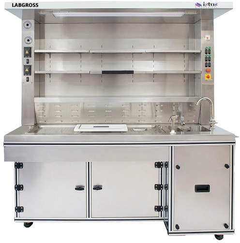
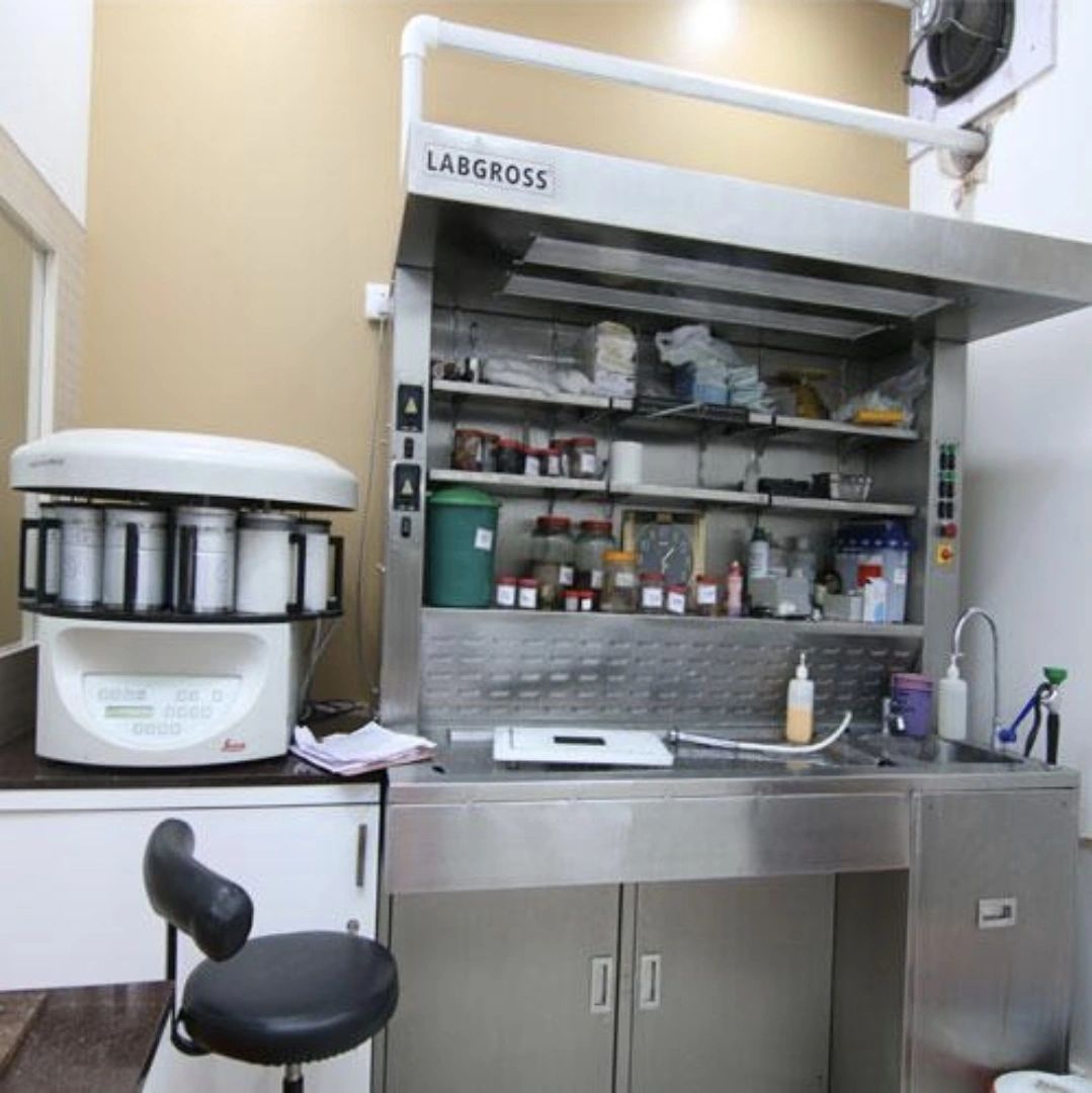
Modern grossing stations, including the ISTOS LABGROSS range, integrate ventilation, splash protection and ergonomic layout so that formalin handling becomes safer and more standardised day‑to‑day.
Conclusion
Used correctly, formaldehyde in 10% Neutral Buffered Formalin underpins almost every routine histology workflow. When fixation time, tissue thickness, safety and ergonomics are all under control, labs gain cleaner morphology, more reliable staining and fewer repeat procedures.
📞 To explore formalin‑handling and grossing solutions tailored to your laboratory, contact ISTOS Medical at +91 80 2686 0607 or +91 99001 98668, or email talktous@istos.in.