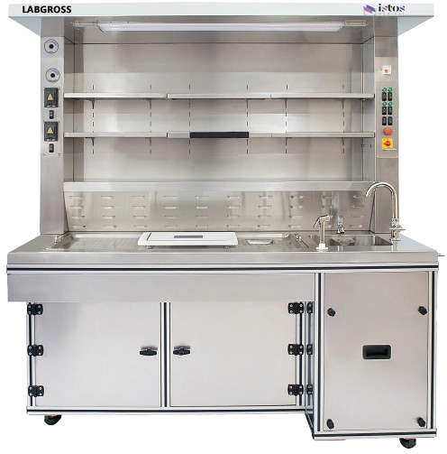
Introduction
Quantitative immunohistochemistry (IHC) has moved beyond a simple “brown or not brown” judgement. Today it is a critical tool for measuring biomarker expression in a reproducible way, especially in oncology and translational research.
Every reported H‑score, percentage of positive cells, or digital readout depends on a quiet assumption: that each section on the slide is comparable to the next. If section thickness, morphology or background staining vary, even the most sophisticated image analysis pipeline will produce unreliable data. That is why ISTOS Medical emphasises section consistency as a formal, documented part of quantitative IHC workflows.
Why section consistency matters
Imagine a biomarker‑driven trial where eligibility depends on at least “50% strong membranous staining” for a particular marker. One site cuts thick, compressed sections; another cuts thin, fragile sections. Both may “follow protocol”, but apparent biomarker expression can shift purely because of microtomy technique rather than underlying biology.
When sectioning is not controlled, labs can face:
- Apparent over‑ or under‑expression because thicker sections trap more chromogen or show more overlapping nuclei.
- Increased background staining or uneven antigen retrieval due to inconsistent tissue depth.
- Poor comparability between timepoints in longitudinal studies or between sites in multi‑centre trials.
- More repeats, more queries from monitors and more difficulty defending the robustness of the dataset.
By contrast, validated, consistent sectioning helps you:
- Maintain uniform staining intensity across slides, so a “3+” today really means the same as a “3+” next month.
- Trust quantitative image analysis outputs because the input sections are standardised.
- Reduce inter‑operator and inter‑site variability, which is crucial when trials run across multiple hospitals or CROs.
- Demonstrate reproducibility when audited or when submitting data to regulatory authorities.
In short, the seemingly routine step of cutting the block can make or break the credibility of quantitative IHC.
Building a section‑consistency validation workflow
ISTOS Medical supports labs not only with instruments but also with practical, repeatable validation workflows. A typical approach to section consistency might include the following elements.
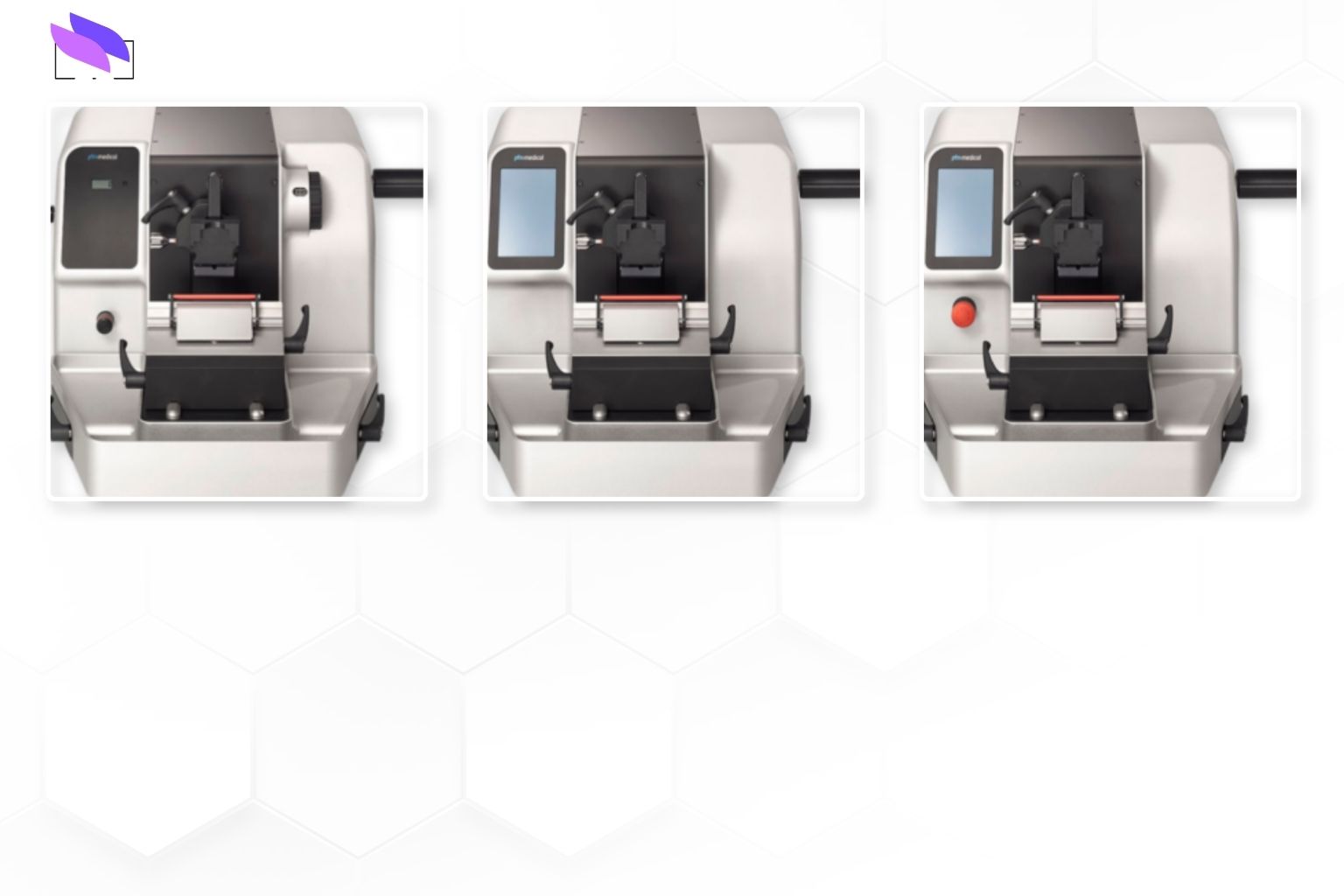
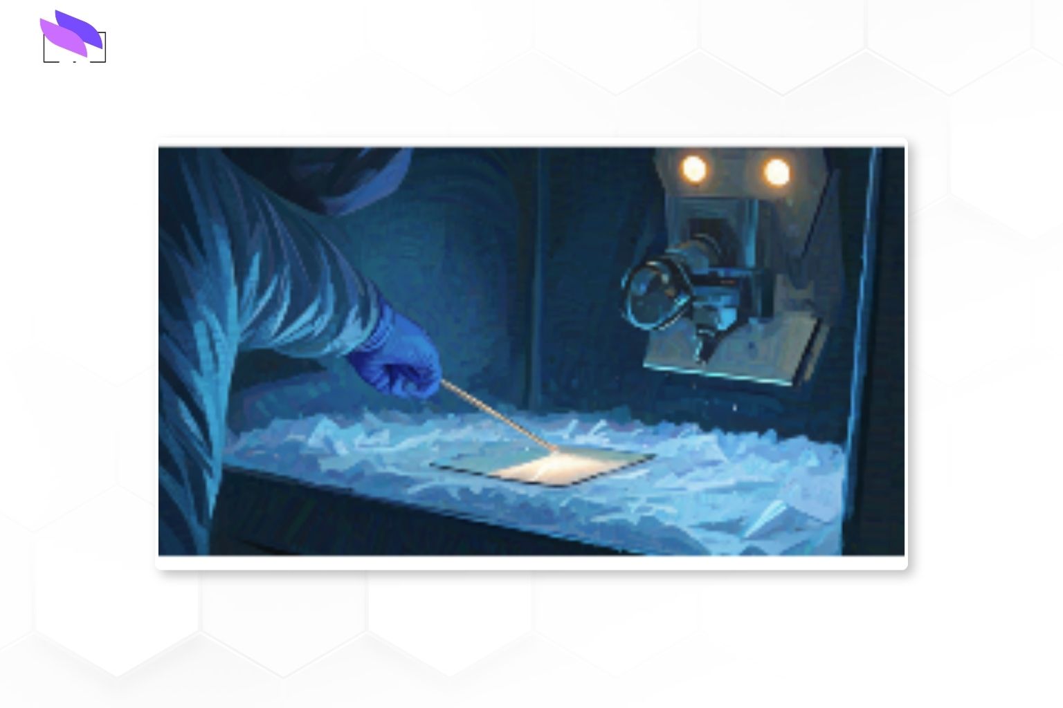
1. Standardising microtomy
The first task is to standardise how sections are cut:
- Define a target thickness range appropriate for IHC, and ensure microtome settings and blade choice support that range reliably.
- Align block trimming, chilling and face‑off procedures so each section starts from a stable, reproducible tissue surface.
- Train staff to recognise artefacts such as chatter, compression, folds and knife marks, and to document when these exceed predefined acceptance thresholds.
Instead of relying solely on individual experience, the process is written down, trained and periodically re‑verified.
2. Using digital checks, not just the naked eye
Once sections are cut and stained, digital tools can help confirm consistency:
- Slide scanners capture high‑resolution images of representative control tissues and study slides.
- Simple quantitative metrics, such as mean optical density in control regions or distributions of staining intensities, are tracked over time.
- Shifts in these metrics often reveal changes in section thickness or cutting quality before they are obvious by eye.
This turns “sections look a bit thick today” into measurable, monitorable data.
3. Leveraging image analysis to monitor drift
Many labs already use image analysis for quantitative IHC. The same platforms can be used to monitor section consistency:
- Compare biomarker expression in standardised control tissues across runs to detect drift.
- Set acceptable ranges for background, noise or signal‑to‑noise ratio that indirectly reflect section quality.
- Flag outliers automatically so problematic runs or blocks are identified early and can be repeated or excluded according to protocol.
The goal is documented control: when a deviation happens, the lab knows, records it and takes appropriate action.
4. Cross‑validating across people and platforms
Even well‑designed processes can drift if only one operator or instrument is ever tested. A robust validation includes:
- Running the same blocks through multiple microtomes and operators to confirm that section quality stays within defined limits.
- Repeating a subset of cases on different days or shifts to capture realistic variation.
- Documenting concordance between runs, including when repeatability thresholds are not met and what corrective actions were taken.
This kind of cross‑validation reassures both internal stakeholders and external reviewers that section quality — and therefore quantitative IHC data — does not depend on a single “star technician” or a single instrument.
Efficiency and data quality go together
Investing time in section consistency validation may look like extra work, but stabilising the process usually makes day‑to‑day operations easier.
- Fewer repeats mean less waste of reagents, slides and technologist time.
- Consistent sections lead to smoother staining, fewer troubleshooting sessions and less back‑and‑forth between histology and pathology.
- Trial sponsors and monitors gain confidence in the data, which helps avoid delays during interim analyses or final submissions.
- Pathologists can focus more on interpretation and less on questioning whether a weak or strong stain is technical or biological.
For research teams, this translates into cleaner datasets, stronger statistical power and more persuasive conclusions when quantitative IHC data are used to support publications or regulatory filings.
How section variability distorts quantitative IHC
Key elements of a section‑consistency validation plan
How section consistency improves both efficiency and data quality
Conclusion
Clinical validation of section consistency turns an often overlooked pre‑analytical step into a controlled, measurable part of your quantitative IHC pipeline. When sections are cut, stained and analysed within a validated framework, biomarker metrics become more trustworthy and the downstream clinical decisions they support become more defensible.
Whether you are building a new trial platform, expanding your IHC menu or upgrading instrumentation, ISTOS Medical can help integrate section consistency validation into your broader quality strategy — so your lab delivers not only beautiful slides but truly reliable numbers.
📞 Contact ISTOS Medical at +91 80 2686 0607 or +91 99001 98668, or email talktous@istos.in to discuss validation workflows, site‑specific implementations and instrumentation for quantitative IHC.